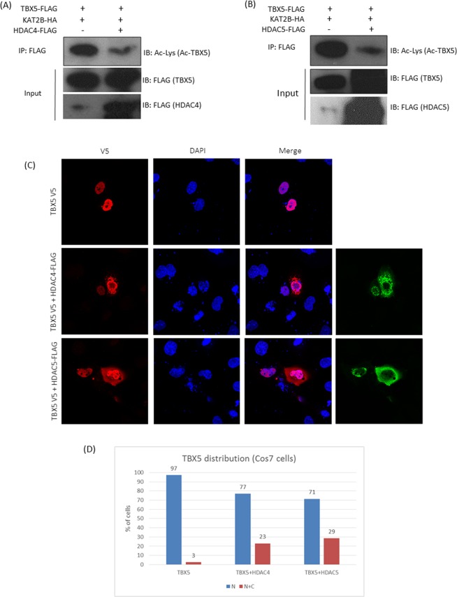
Figure 2.
HDAC4 and HDAC5 deacetylate TBX5 and promote its nuclear export. (A,B) Pull-down and Western blot analysis showing that both HDAC4 and HDAC5 strongly deacetylate TBX5. KAT2B was used to promote TBX5 acetylation. Full-length blots are shown in Supplementary Fig. S2 (A–D). (C) Representative images of the cellular distribution of TBX5 following HDAC4/HDAC5 overexpression, showing the partial re-localization of TBX5 into the cytoplasm. (D) Cell count showing the cellular distribution of TBX5 in cells transfected with TBX5 alone or TBX5 and HDAC4/5 (TBX5- total counts 300; 292 (N) and 8 (N + C), TBX5 + HDAC4- total cell counts 257; 198(N) and 59 (N + C) and TBX5 + HDAC5− total counts 274; 195(N) and 79(N + C). Results are from three individual experiments (N = nuclear, C = cytoplasmic).

