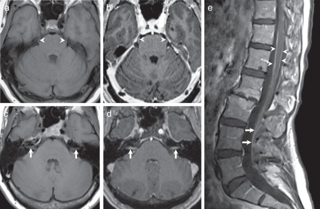
Fig. 1.
MRI of the brain and spine with and without contrast at diagnosis. a, b T1-weighted axial images show enhancement of the bilateral fifth cranial nerves after the administration of gadolinium contrast. c, d There is enhancement of the bilateral seventh–eighth cranial nerve complexes. e Sagittal T1-weighted post-contrast spine images demonstrate patchy circumferential enhancement along the thoracic and lumbar spinal cord (arrow heads) and enhancement of the cauda equina nerve roots (arrows), related to leptomeningeal carcinomatosis.

