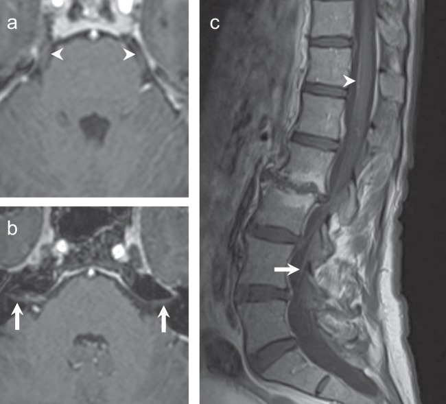
Fig. 2.
MRI brain and spine, 12 months following treatment with olaparib. a, b Axial T1-weighted post-contrast images show resolved enhancement of the fifth cranial nerves (a, arrowhead) and seventh–eighth cranial nerves complexes (b, arrows). Linear enhancement adjacent to the right seventh–eighth cranial nerve complex was consistent with a vascular loop. c Sagittal T1-weight MRI of the spine, post contrast, shows minimal enhancement anterior to the thoracic spinal cord (arrowhead) and subtle enhancement within the cauda equina nerve roots (arrow).

