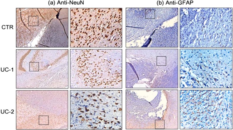
Figure 8.
Trial 2 Immunohistochemistry (Day 5 Treatment). IHC staining of brain samples from Untreated Control (CTR) (top row), UC-1- (middle row), and UC-2-treated rats (bottom row) that died spontaneously from 9L gliosarcoma at days 12, 43, and 30, respectively. For each pair of images, left is 2.5× magnification of the area at the margin between tumor and healthy tissue and right is 20× magnification of healthy tissue in proximity to tumor margin (general location demonstrated by square boxes in 2.5× photos). Anti-NeuN (a) and anti-GFAP (b) staining demonstrates no qualitative decrease in signal from healthy neurons and astrocytes among treated brains compared to control. Images of astrocytes and neurons were obtained from hippocampal and cortical areas, adjacent to the tumor margin near wafer implant.

