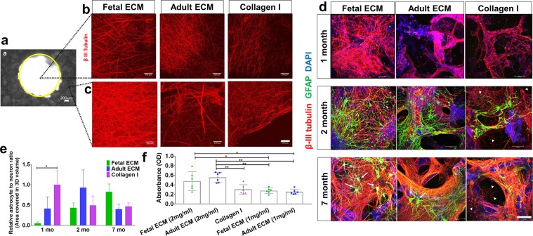
Figure 2.
Extracellular matrix and time-dependent differentiation of human induced neural stem cells in 3D cultures. Human induced neural stem cells (hiNSCs) in silk scaffold-based 3D constructs infused with collagen I hydrogels supplemented with native porcine brain-derived ECM. (a) Brightfield image of silk scaffold with the middle circular window indicated by the yellow outline. (b) Growth of differentiating hiNSCs at 6 wk. shown by β-III tubulin staining for neurons within the middle hydrogel window of the 3D donut-shaped constructs. Max projection of z-stack. Scale bar 100 μm. (c) Growth of differentiating hiNSCs at 6 wk. shown by β-III tubulin staining for neurons within the ring portion of the 3D donut-shaped constructs. Max projection of z-stack. Scale bar 100 μm. (d) Growth and differentiation of hiNSCs at 1, 2 and 7 mo. shown by β-III tubulin staining for neurons (red) and GFAP staining for astrocytes (green) across different ECM conditions. Max projection of z-stack. Scale bar 100 μm. Arrows point to the star shaped astrocytes and arrow heads to the disintegrated axons and neuronal debris. (e) Astrocyte to neuron ratio calculated by dividing the total volume in 3D confocal stacks covered by astrocytes versus neurons post image processing. Mean ± SEM, One-way ANOVA with Dunnett’s post hoc test (Collagen I as control condition) at each time point on log transformed data, n = 3–6 individual scaffolds per condition. (f) Wst-1 viability assay at 2.5 mo. in 3D hiNSC cultures. One-way ANOVA with Tukey’s post hoc for multiple comparisons. *p < 0.0431, **p < 0.0071.

