Figure 1.
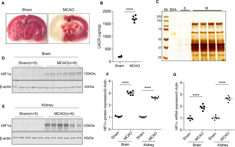
Ischemic stroke alters kidney function. (A) TTC staining images of sham and ischemic stroke-induced (MCAO) rat brain. (B) Estimation of albumin and creatinine levels in MCAO rats. Error bars indicate mean ± SE; n = 6. ****p < 0.0001. (C) Urine samples from sham (S) and stroke-induced rats (M) were subjected to SDS-PAGE and urinary proteins were visualized by silver staining, Mr, molecular weight marker (#1610374; Bio-Rad); BSA, Bovine serum albumin. HIF1α expression in the infarcted region of the brain (D) and glomerular lysates (E) from sham and MCAO rats. Densitometric analysis of HIF1α band is depicted after normalized for respective β-actin expression (F). Error bars indicate mean ± SE; n = 5–6. ****p < 0.0001. (G) Steady-state mRNA levels of HIF1α from the brain and glomerular lysates were measured by qRT-PCR. Error bars indicate mean ± SE; n = 6. ****p < 0.0001.

