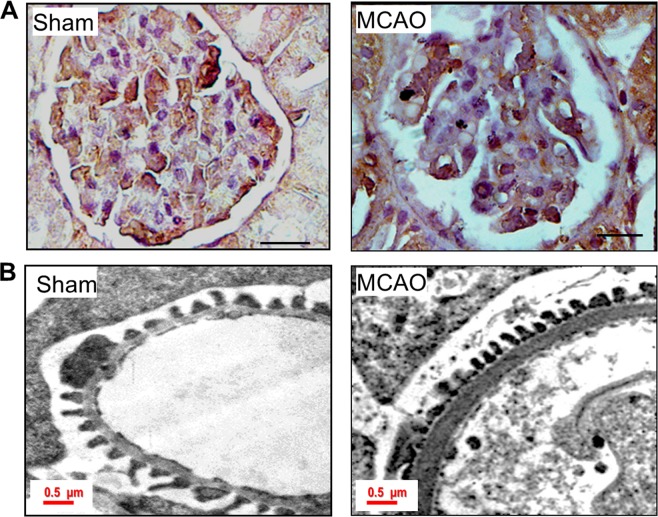
Figure 2.
Ischemic-hypoxia elicits podocyte injury. (A) Staining for WT1 in sham and MCAO rat glomerular sections. The scale bar represents images of 10 µm and images were captured with a 100x objective of Leica trinocular microscope. (B) TEM images of podocyte foot processes in sham and MCAO rat kidney sections. In ischemic-stroke rats, podocyte foot-processes were small and the thickness of GBM was increased compared to sham-operated rats.

