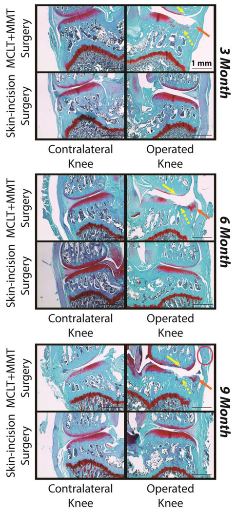
Figure 4: Representative Histological Images.
Images of the medial compartment are provided for the operated knee (right) and contralateral knee (left). In all age groups, tibial cartilage damage was observed (yellow, dashed arrows), femoral cartilage damage was observed (yellow, solid arrows), and osteophyte formation was observed (orange arrows). Synovitis was apparent is some samples from MCLT+MMT operated knees (pink oval), but was not consistently observed at any age group.

