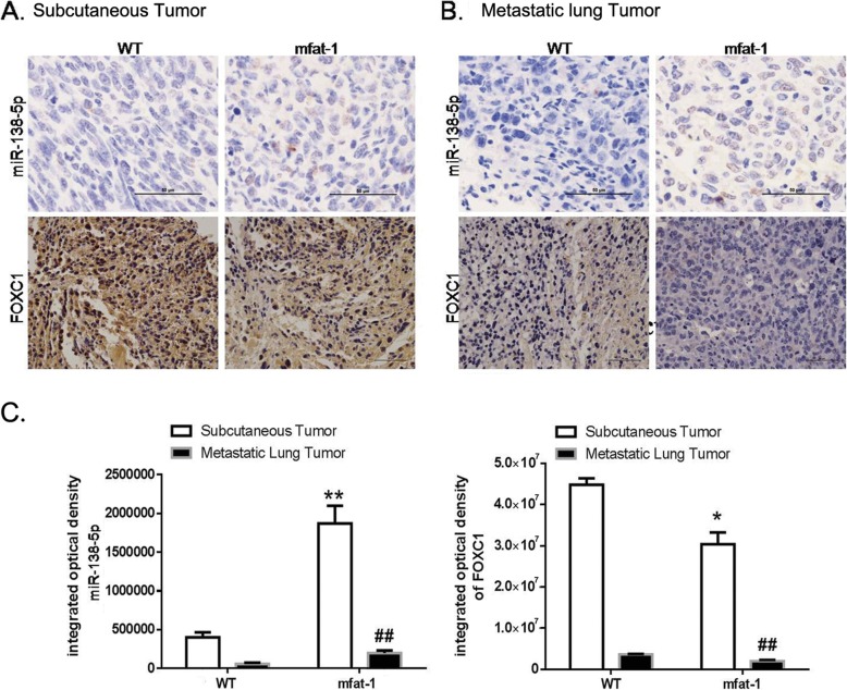
Fig. 6.
Endogenous DHA increases miR-138-5p expression and decreases FOXC1 expression in vivo. LLC cells were injected subcutaneously into WT or mfat-1 mice (a), or injected into the tail vein of WT or mfat-1 mice (b). Representative in situ hybridization images of cancer tissues stained with miR-138-5p RNAscope probe (upper panel). Representative immunohistochemical images of LUSC tissues and LUAC tissues stained with an anti-FOXC1 antibody (lower panel). Scale bars = 50 μm (40×). c. The integrated optical density level was determined using Image Pro Plus software. Data are presented as the mean ± SEM from 4 different samples. *P < 0.05, **P < 0.01 for miR-138-5p expression compared to WT group; ##P < 0.01 for FOXC1 expression compared to WT group

