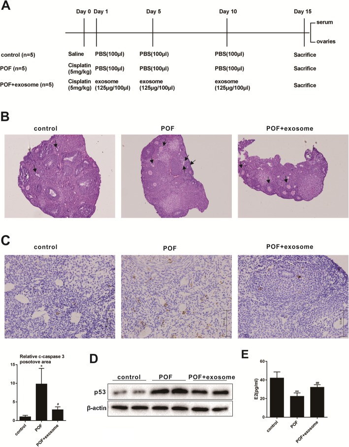
Fig. 2.
Therapeutic effect of BMSC-derived exosomes on POF. a Protocol for mouse model of POF. b HE staining of ovarian tissue was performed to observe the morphological differences of follicles (under (× 100) magnification, arrows point to atretic follicles). c The difference in the expression of cleaved-caspase3 in the ovarian tissue was analyzed by immunohistochemistry (scale bar, 5.0 μm) *P<0.05 vs.control group, #P<0.05 vs. POF group. d The difference expression of P53 in the ovarian tissue was detected by Western blot. e The concentration of E2 in the serum was detected by ELISA **P<0.01 vs.control group, ##P<0.01vs. POF group. Data are expressed as mean ± standard deviation (SD)

