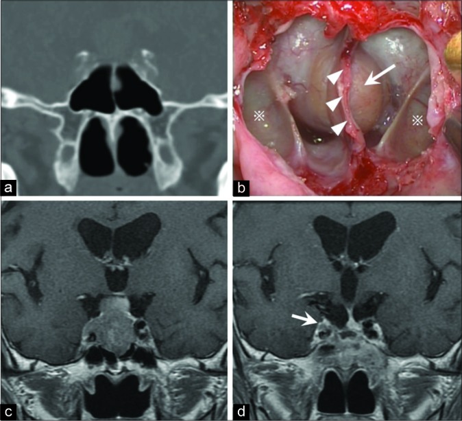
Figure 2:

Illustrative case with images and operation view. (a) Preoperative computed tomography in a coronal section showing the developed lateral recess of the sphenoid sinus (LRSS). (b) This intraoperative view shows the posterior wall of the SS from an endoscopic endonasal transsphenoidal approach, including the LRSS (※), septum of the SS (head arrow), and sella turcica (arrow). (c) Preoperative gadolinium-enhanced T1-weighted magnetic resonance imaging (Gd-T1WI MRI) in a coronal section showing a pituitary adenoma with cavernous sinus (CS) invasion. (d) Postoperative Gd-T1WI MRI showing that almost all of the tumor was removed but there was a small residual tumor on the internal carotid artery in the right CS (arrow).
