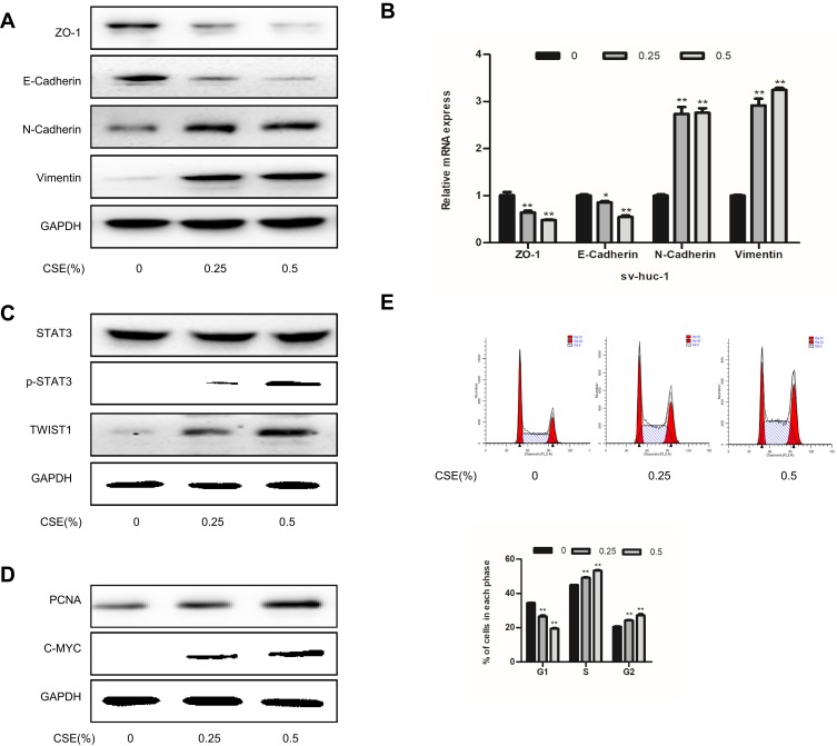
Figure 2.
CSE exposure induces EMT in SV-HUC-1 cells. (A) Western blotting detected the protein expression of epithelial markers and mesenchymal markers. (B) The mRNA expression level of SV-HUC-1 cells following CSE treatment was measured by qRT-PCR. (C) Western blotting reveals the expression of STAT3 phosphorylation in the SV-HUC-1 cells following CSE treatment for 5 days. (D) The protein expression of cell proliferation protein was measured by Western blotting. (E) Cell cycle was detected by flow cytometry. Data are expressed as mean ± SD. The values of *P<0.05, **P<0.01 show a statistical level of significance when data were compared against the control group.

