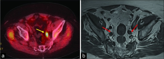
Figure 1:

An 85-year-old male with history of prostate cancer status post radiation treatment presented with rising prostate- specific antigen level of 6.1 ng/mL. (a) Axumin positron emission tomography-computed tomography axial image showing increased radiotracer uptake (standardized uptake value maximum of 5.3) in the left internal iliac lymph node (yellow arrow). (b) Magnetic resonance imaging pelvis T1 weighted axial image depicting a few enlarged bilateral internal iliac lymph nodes (red arrow) which were otherwise inconclusive for recurrence.
