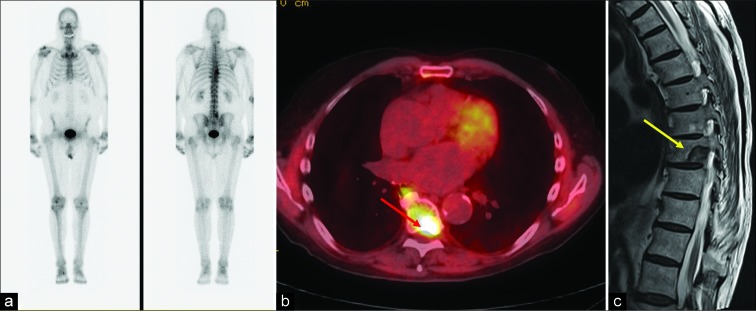
Figure 3:
An 88-year-old male with history of prostate cancer status post prostatectomy presented with progressive back pain and prostate- specific antigen level of 6.9 ng/mL concerning for recurrence. (a) Bone scan was negative. (b) Axumin positron emission tomography- computed tomography axial image demonstrating intense tracer uptake (standardized uptake value maximum 6.4) in the left posterolateral aspect of the T8 vertebral body (red arrow). (c) Pre-biopsy magnetic resonance imaging performed showing T2 hypointense lesion (yellow arrow) measuring 2.2 cm which was consistent with osteoblastic metastasis on biopsy.

