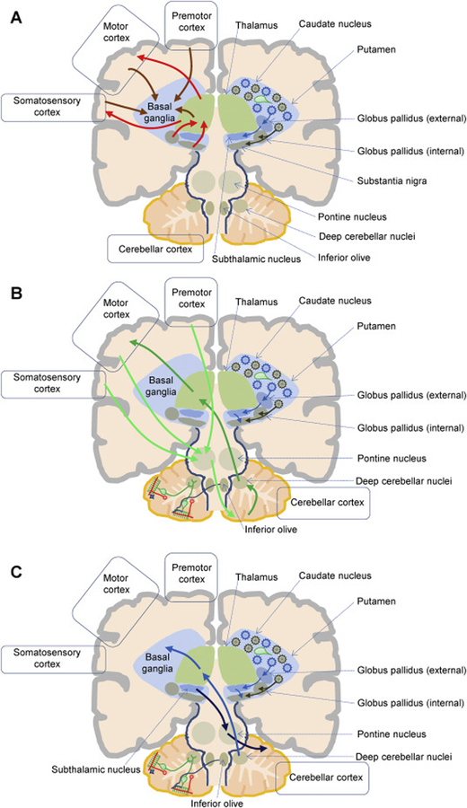
Figure 1. Simplified cartoon of basal ganglia-cerebellum anatomical connections.
A. The cartoon depicts a simplified cortico-basal ganglia-thalamo-cortical circuit. Afferents from the cortex and thalamus (left, orange arrows) reach caudate and putamen neuronal populations of cholinergic interneurons (right, green) and projection neurons. Spiny projection neurons give rise to the direct (right, brown) and indirect (right, blue) output pathways, ultimately reaching the basal ganglia output nuclei (globus pallidus externus and substantia nigra pars reticulata), which send the processed information to the thalamus (left, red arrows). The thalamus then project back to the cortex (left, red arrow). B. The cartoon depicts simplified cortico-cerebello-thalamo-cortical connections. Left. The cerebellar cortex has a trilaminar cytoarchitecture composed of the Purkinje cell layer and the molecular layer (green), and of the granular layer (granule cells, red). Parallel fibers (red), originating from granule cells, reach the molecular layer and innervate inhibitory stellate (blue) and basket interneurons, as well as Purkinje cells (green). Purkinje cells are also activated by climbing fibers (blue), which originate in the inferior olive. The pontine nucleus receives cortical afferents and is a source of mossy fibers reaching the contralateral cerebellar cortex (light green arrow). Purkinje cells project to the deep cerebellar nuclei, which in turn convey cerebellar output to the cortex via the thalamus (green arrows). C. Retrograde transneuronal transport of rabies virus in monkeys revealed bidirectional disynaptic connections between the cerebellum and the basal ganglia. Specifically, a pathway originating from the deep cerebellar nuclei and reaching the putamen (blue arrows; Hoshi et al., 2005), and a pathway connecting the subthalamic nucleus to the cerebellar cortex (light blue arrows; Bostan et al., 2010) were identified.

