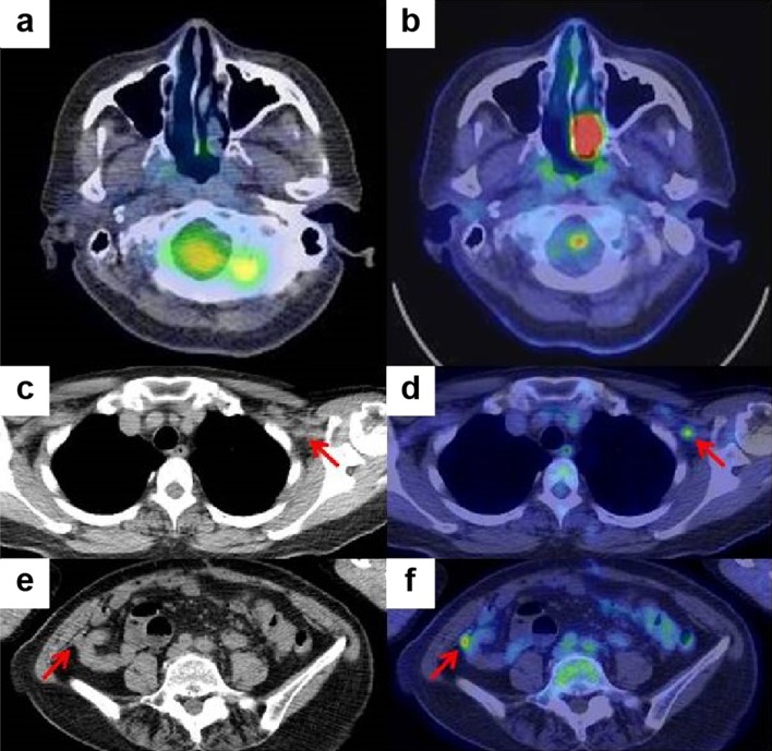
Fig. 8.
Positron emission tomography–computed tomography image of the nasal cavity before (a) and after surgery (b). Plane computed tomography and positron emission tomography–computed tomography images after surgery revealed left infraclavicular lymph node metastasis (c, d) and a small peritoneal nodule (e, f)

