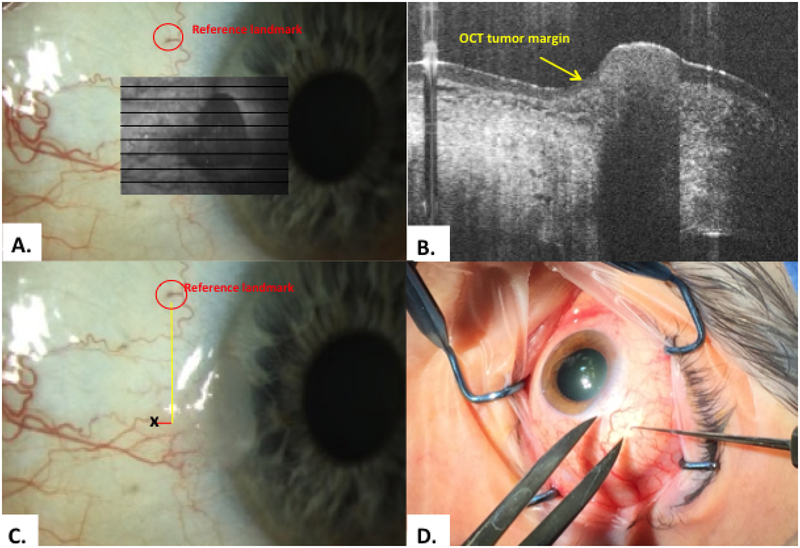
Figure 2.
Methodology: scanning, and transfer of HR-OCT predicted tumor margin points A. A reference point was identified of a prominent vessel or marking (red circle). The tumor was scanned to identify lesion margins. The en-face overlay is shown here in the gray box. B. Classic OCT findings of epithelial hyper-reflectivity and abrupt transition from normal to abnormal were identified (yellow arrow). C. The internal calipers of the OCT device measured the distance from the reference point to the HR-OCT predicted tumor margin (black x). D. Example of transfer method of OCT predicted tumor margin points intra-operatively using calipers and a marked hook.

