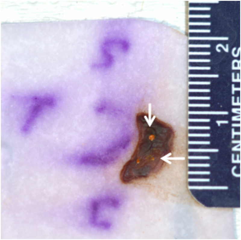
Figure 3:
Patient 1 conjunctival tumor specimen
Following excision, tumor was placed on paper and oriented, superior (S), temporal (T) and inferior (I). The transferred HR-OCT predicted margins were re-marked with permanent orange dye prior to fixation. Note the variability in the size of the re-marked orange ink dots with one being prominent and the other two quite scant (arrows).

