Figure 6.
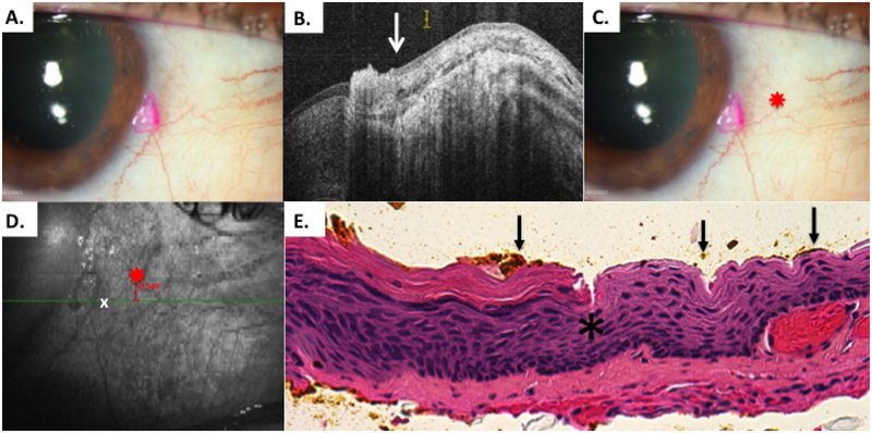
Patient 3 conjunctival tumor with leukoplakia
A) Slit lamp photo of right eye from patient 3 with a discreet, leukoplakic lesion consistent with OSSN. B) HR-OCT cut with predicted tumor margin denoted by white arrow. Note thickening and strong hyper-reflectivity over tumor. Adjacent bulbar conjunctiva has normal thickness but remains somewhat hyper-reflective epithelium adjacent to tumor, likely secondary to actinic changes. C) Slit lamp photo of right eye showing selected reference landmark (red star). D) OCT en-face reconstruction of ocular surface with reference landmark denoted by red star and measurement to HR-OCT predicted tumor margin (white x) with internal calipers E) Examination of the excised conjunctiva discloses faulty epithelial maturational sequencing that extends up to full thickness with overlying hyperkeratosis (carcinoma in situ) the area of predicted tumor margin (asterisk) is present adjacent to the transition of variably acanthotic and dysplastic epithelium. OCT predicted mark (orange pigment, black arrows) coinciding with pathologically identified margin (Hematoxylin-eosin; original magnification x400).

