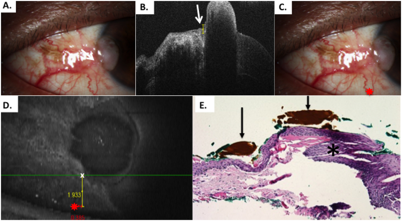
Figure 8.
Patient 6 corneal and conjunctival tumor
A) Slit lamp photo of diffuse conjunctival OSSN with opalescent corneal component. Note conjunctival melanosis in this patient with dark complexion. B) HR-OCT cut with predicted tumor margin denoted by arrow. C) Slit lamp photo of right eye showing selected reference landmark (red star) of looped vessel. D) OCT en-face reconstruction of ocular surface with reference landmark denoted by red star and measurement of inferior HR-OCT predicted tumor margin (white x) from reference landmark E) Examination of the excised, tangentially sectioned conjunctiva discloses faulty epithelial maturational sequencing that extends up to full thickness (carcinoma in situ) with the area of predicted tumor margin (arrows; orange pigment) adjacent to the transition of unremarkable and dysplastic epithelium (asterisk) (Hematoxylin-eosin; original magnification x200).

