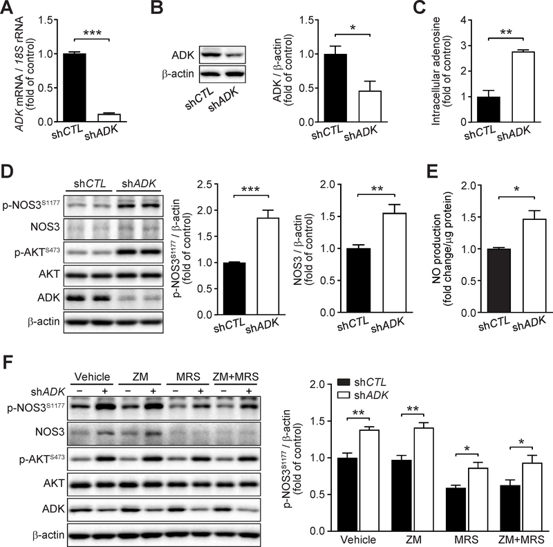
Figure 7. Increased endothelial nitric oxide synthase (NOS3)/nitic oxide (NO) pathway in ADK knockdown endothelial cells.
A. qPCR analysis of adenosine kinase (ADK) expression in human umbilical vein endothelial cells (HUVECs) infected with control shRNA (shCTL) or ADK shRNA (shADK) adenovirus for 48 h (n = 6). B. Representative Western blot results of ADK and β-actin in HUVECs infected with shCTL or shADK adenovirus for 48 h (left) and relative ratio of ADK/β-actin (right) were quantitated by densitometric analysis of the corresponding Western blots (n = 3). C. Quantification of relative intracellular adenosine concentration in control (shCTL) and ADK knockdown (shADK) HUVECs (n = 3). D. Representative Western blot results of phospho-NOS3 (Ser1177) (p-NOS3S1177), total NOS3 (NOS3), phospho-AKT (Ser473) (p-AKTS473), total AKT (AKT), ADK and β-actin in HUVECs infected with shCTL or shADK adenovirus for 48 h (left) and relative ratio of p-NOS3S1177/β-actin (middle) and NOS3/β-actin (right) were quantitated by densitometric analysis of the corresponding Western blots (n = 6). E. Quantification of relative NO concentration in the culture medium of HUVECs infected with control shRNA (shCTL) or ADK shRNA (shADK) adenovirus for 48 h (n = 4). F. Representative Western blot results of endothelial phospho-NOS3 (Ser1177) (p-NOS3S1177), total NOS3 (NOS3), phospho-AKT (Ser473) (p-AKTS473), total AKT (AKT), ADK and β-actin (left). HUVECs were infected with shCTL or shADK adenovirus for 24 h, then treated with ZM 241385 (5 μM), MRS 1754 (5 μM) or both ZM 241385 and MRS 1754 for another 24 h. Relative ratio of p- NOS3S1177/β-actin (right) were quantitated by densitometric analysis of the corresponding Western blots (n = 4). All data are represented as mean ± SEM, *P < 0.05, **P < 0.01 and ***P < 0.001 for indicated comparisons (unpaired two-tailed Student’s t test).

