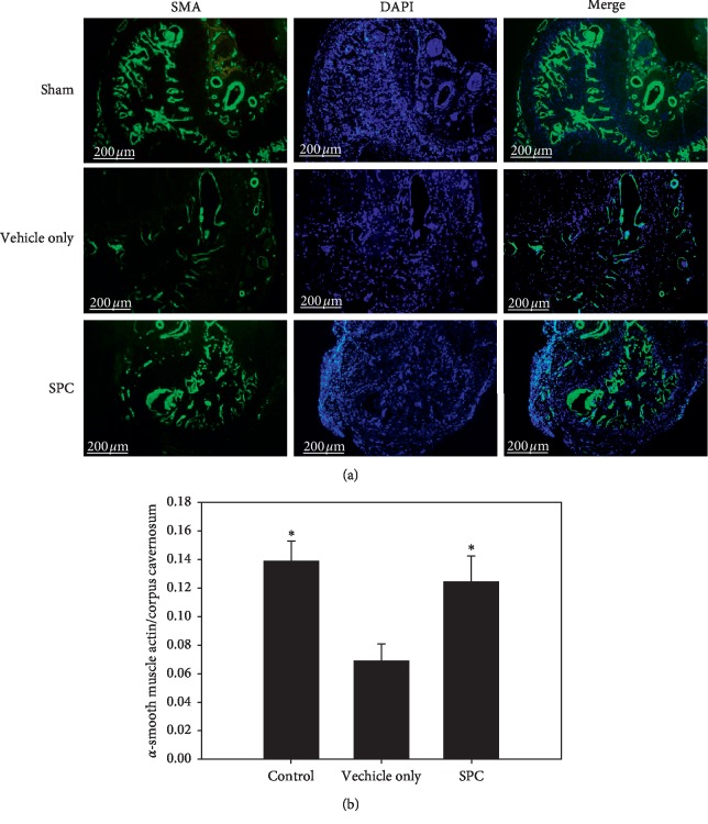
Figure 4.
Immunofluorescence staining of α-SMA in smooth muscle cell of corpus cavernosum. Histological analyses of the corpus cavernosum (CC) 28 days after injury. (a) Representative fluorescence images of α-SMA-positive areas in the rat penile CC (smooth muscle, green; nuclei, blue) (original magnification: 50×). (b) Graph showing the smooth muscle cell (SMC) content in the CC quantified as the α-SMA-positive area/CC area. ∗p < 0.05 versus the vehicle group.

