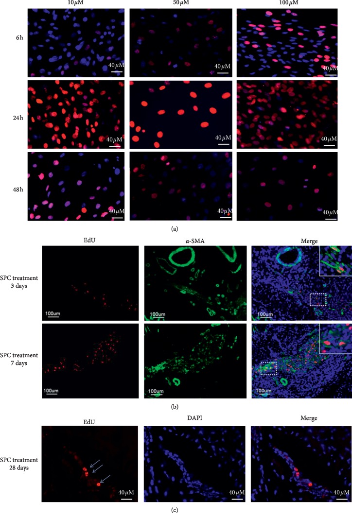
Figure 6.
EdU labeling of smooth muscle progenitor cells (SPCs). (a) SPCs labeled with EdU at 10, 50, and 100 μM and stained with Alexa-594 (red fluorescence) and DAPI (blue fluorescence) (200x magnification). Tracking corpus cavernosum-transplanted SPCs. (b, c) SPCs were labeled with EdU and injected into the corpus cavernosum tissue, which was then harvested at 3, 7, and 28 d and stained with Alexa-594 (red fluorescence) and DAPI (blue fluorescence). The Alexa-594 and DAPI stained images were digitally merged (scale bar = 40 μm and 100 μm).

