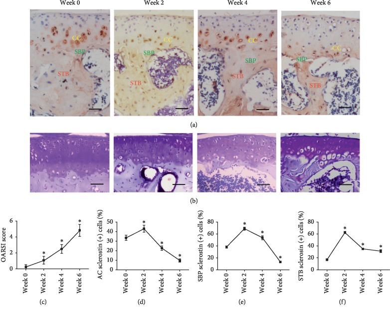
Figure 2.
Expression trend of sclerostin in the CC, SBP, and STB of mice after ACLT. (a) The immunohistochemical staining of sclerostin in CC, SBP, and STB. CC: calcified cartilage; SBP: subchondral bone plate; STB: subchondral trabecular bone. (b) The toluidine blue staining revealed that OA in the tibial plateau was time dependently aggravated after ACLT in mice. Bar, 100 µm. (c) OARSI scores of knee samples increased overtime after ACLT. (d–f) The percentage of sclerostin-positive cells in the CC, SBP, and STB. The expression of sclerostin in the CC, SBP, and STB exhibited the same trend, which increased from week zero to week two and decreased from week two to week six (∗P < 0.05vs. week zero; the data were presented as mean ± standard deviation (SD)).

