. 2019 Nov 3;16(6):505–518. doi: 10.21873/cgp.20153

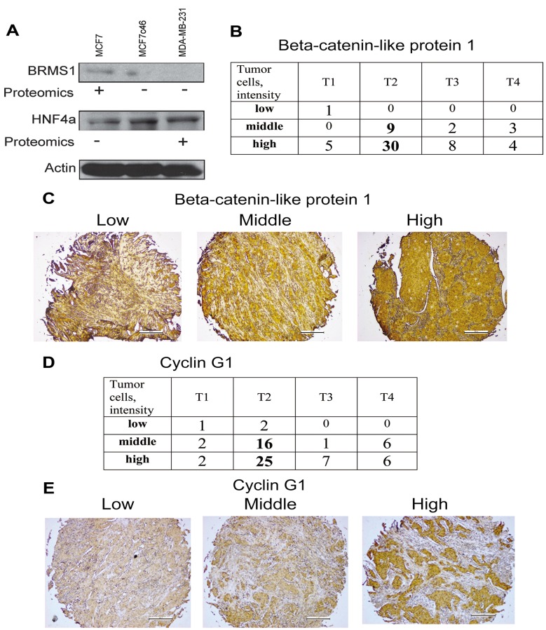
Figure 2. Validation studies confirmed the proteomic data. A) Expression of BRMS1 and HNF4α in MCF7, MCF7c46 and MDA-MB-231 cells. Immunoblotting confirmed expression pattern of these proteins detected in the proteome profiling. For proteomics entries for BRMS1 see #64 in Supplementary Table I and #157 in the Supplementary Table II, and for HNF4α see #51. (+) indicates increased expression. Actin immunoblotting was performed for control of loading. B) Immunohistochemistry of β-catenin-like protein 1. Tissue microarray of 62 cases of invasive ductal carcinomas was used for evaluation of β-catenin-like protein 1 expression. Intensity of staining of tumor cells (low, middle and high) in relation to T1, T2, T3 and T4 of TNM classification of cases is presented as numbers of cases. C) Representative images of low (left), middle (middle) and high (right) staining of tissues for expression of β-catenin-like protein 1 are shown. D) Immunohistochemistry of cyclin G1. Tissue microarray of 68 cases of invasive ductal carcinomas was used for evaluation of cyclin G1 expression. Intensity of staining of tumor cells (low, middle and high) in relation to T1, T2, T3 and T4 of TNM classification of cases is presented as numbers of cases. E) Representative images of low (left), middle (middle) and high (right) staining of tissues for expression of cyclin G1 are shown.

Figure 2