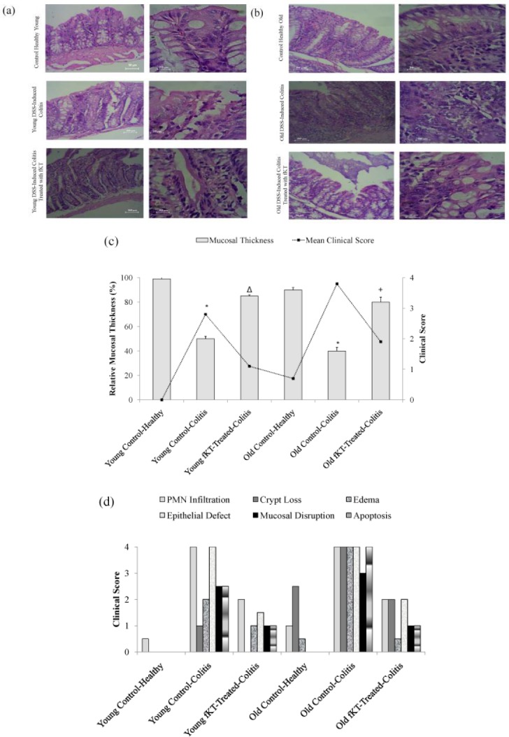
Figure 6.

(a) H&E stained colon sections of healthy young animals, young DSS-induced colitis, and young DSS-induced colitis treated with fKT, (b) healthy old animals, old DSS-induced colitis, and old DSS-induced colitis treated with fKT comparing PMNs, cryptic loss, epithelial defect, mucosal disruption, apoptosis, edema, and mucosal thickness. Colitis was induced by administration of 3.5% DSS in drinking water for seven days, and treatment with fKT was performed for the next seven days. The picture was taken using an Olympus D330 digital camera (Olympus, Tokyo, Japan). The right panel with a magnification of x10 and the left with ×40. (c) Thickness changes in the colon of healthy young animals, young DSS-induced colitis, and young DSS-induced colitis treated with fKT, healthy old animals, old DSS-induced colitis, and old DSS-induced colitis treated with fKT. The thickness of mucosa colon was estimated relative to age-matched healthy animals in a total of six areas per sample with fixed interval (*P< 0.05 vs control, Δ & +P<0.01 vs DSS-mice). (d) Damage score ranged from 0 to 4, scales judged based on the number and extent of PMN infiltration, epithelial defects, crypt loss, mucosal disruption, edema, and apoptosis, as described in the text
