Figure 4.

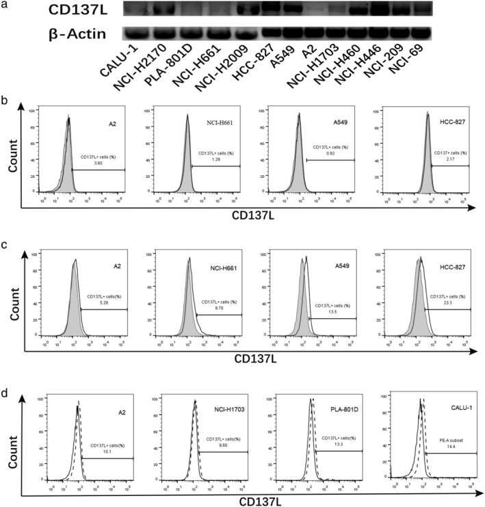
CD137L expression levels were detected by RT‐PCR and flow cytometry with staining directly or indirectly compared to isotype. (a) RT‐PCR analysis of lung cancer cell lines CD137L mRNA expression. (b) Lung cancer cell lines were each stained with mouse antihuman CD137L mAb (open histograms) or mouse IgG1, κ isotype control (shaded histograms). (c) The cells were stained with human CD137 protein (with the Fc region of human IgG) (open histograms) or human B7‐2Ig (shaded histograms), followed by staining with a secondary rabbit antihuman IgG polyclonal Ab. (d) The cells were treated with IFN‐γ (dotted line) and control (open histograms) to analyze the CD137L expression by flow cytometry.  CD137L,
  Isotype,
  Control and
  IFN‐γ.
