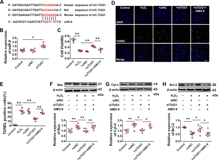
Fig. 2. Silencing lncR-TUG1 alleviates H2O2-induced cardiomyocytes apoptosis by targeting miR-9.
a The bioinformatics analysis showing the binding site for miR-9 with lncR-TUG1. b Increase in miR-9 level after LncR-TUG1 knockdown by siRNA (n = 6). *P < 0.05 by Student’s t-test. Data are presented as mean ± SEM. NRVMs were divided into five groups: control, H2O2 treatment, H2O2 + siNC, H2O2 + siTUG1, and H2O2 + siTUG1 + AMO-9. c Alterations of cell viability of NRVMs by MTT assay (n = 5). **P < 0.01 by one-way ANOVA analysis with Tukey’s multiple comparison test. Data are presented as mean ± SEM. d Representative images of TUNEL staining of NRVMs for DNA defragmentation showing the apoptotic cells (scale bar: 60 μm). e Statistical results of TUNEL-positive cells per field (n = 5). **P < 0.01 by one-way ANOVA analysis with Tukey’s multiple comparison test. Data are presented as mean ± SEM. f–h Western blot analysis of protein levels of Bax (n = 6), cytochrome-c (Cyt-c; n = 5), and Bcl-2 (n = 7) in NRVMs with different treatments. *P < 0.05, **P < 0.01 by one-way ANOVA analysis with Tukey’s multiple comparison test. Data are presented as mean ± SEM.

