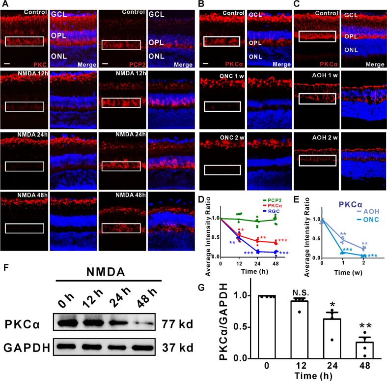
Fig. 1. PKCα degradation in RBCs dendrites appeared to be a universal mechanism for RGCs loss in experimental glaucoma models.
a PKCα expression at the somas and dendrites of RBCs was reduced dramatically after intravitreal injection of NMDA (left), while PCP2 expression remained the same with NMDA treatment (right). b Decreased PKCα expression in RBCs dendrites induced by optic nerve crush (ONC) model. c Representative images of PKCα (red) of retinas treated with the acute ocular hypertension (AOH) glaucoma model. Decreased PKCα expression in RBCs dendrites, and a reduction of the thickness of the retina at time points following administration of NMDA. d, e Fluorescence intensity of PKCα, PCP2, and DAPI in OPL decreased after NMDA treatment. PKCα average intensity ratio of NMDA 12, 24, and 48 h, n = 5: 0.56 ± 0.06, P = 0.0149; 0.41 ± 0.04, P = 0.0013; 0.38 ± 0.02, P = 0.0002. f, g The PKCα protein expression level decreased significantly after the administration of NMDA. Average intensity of PKCα/GAPDH of NMDA 12, 24, and 48 h, n = 4: 0.91 ± 0.05, P = 0.1822; 0.63 ± 0.11, P = 0.0396; 0.26 ± 0.08, P = 0.0029. INL, inner nuclear layer; GCL, ganglion cell layer; OPL: outer plexiform layer. Scale bar = 50 μm.

