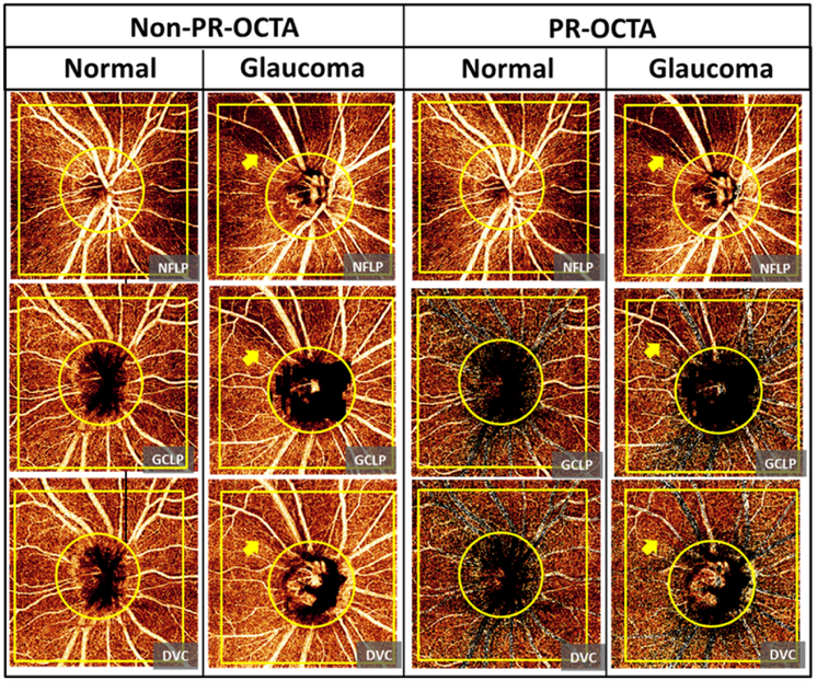
Figure 3.
Comparison of the ganglion cell layer plexus (GCLP) and the deep vascular complex (DVC) angiograms between non projection-resolved optical coherence tomography angiography (non-PR-OCTA) and PR-OCTA from a normal eye and a perimetric glaucoma eye. The vessel pattern in the nerve fiber layer plexus (NFLP), large vessels and radial capillaries, were projected in the non-PR-OCTA GCLP and DVC angiograms in both normal and glaucoma eyes. PR-OCTA removed these projected patterns in the GCLP and DVC angiograms. The non-PR-OCTA DVC angiogram showed projected perfusion defects (arrow) in the glaucoma eye. The PR-OCTA showed that the DVC was unaffected by glaucoma.

