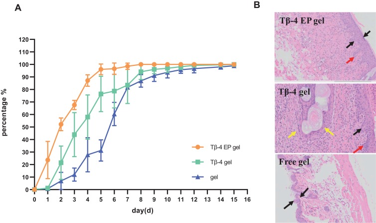
Figure 5.
Kunming mice administrated with different gel for 15 days have different degrees of wound healing (A) and the photomicrographs of the healed skin structure of using different preparations (B). In Figure 5B, the black arow indicated the degree of skin thickening, the red arow indicated new capillaries and the yellow arrow showed embolization and localized parakeratosis.
Abbreviation: Tβ-4 EP gel, Tβ-4 ethosomal gel.

