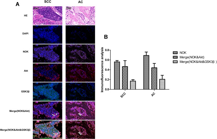
Figure 3.
Co-localization of NOK and Akt and GSK-3β. (SCC, squamous cell carcinoma; AC, adenocarcinoma). (A) HE staining; Immunofluorescence: NSCLC tissue stained with anti-Akt antibody/CY3 (red) and anti-GSK-3β/FITC (green) and anti-NOK antibody/CY5 (pink), and DAPI nuclear stain (blue). (B) Statistics analysis of the co-location of NOK, Akt and GSK-3β. White arrow point to cells with co-localization of NOK & Akt & GSK-3β.

