Figure 3.
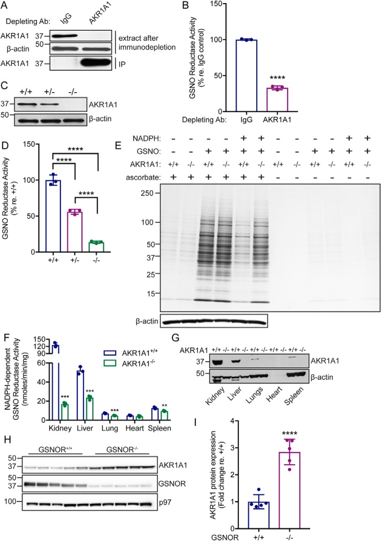
AKR1A1 is an NADPH-dependent GSNO reductase in mammals. A, representative Western blotting analysis following immunodepletion of AKR1A1 from WT mouse kidney extracts. IP, immunoprecipitate. B, relative NADPH-dependent GSNO reductase activity in IgG or AKR1A1 immunodepleted kidney extracts from A. The bars represent means ± S.D. for n = 3. ****, p < 0.0001 by Student's t test. C, representative Western blotting analysis of kidney extracts from AKR1A1+/+, AKR1A1+/−, and AKR1A1−/− mice. The example shown is representative of three independent experiments. D, relative NADPH-dependent GSNO reductase activity in kidney extracts from AKR1A1+/+, AKR1A1+/−, and AKR1A1−/− mice. The bars represent means ± S.D. for n = 3. ****, p < 0.0001 by one-way analysis of variance with Tukey's correction for multiple comparisons. E, representative Coomassie-stained SDS-PAGE gel illustrating SNO-proteins isolated by SNO-RAC following treatment of kidney extracts from AKR1A1+/+ and AKR1A1−/− mice with 0.1 mm GNSO (in the presence or absence of 0.1 mm NADPH). The results are representative of three independent experiments. F, NADPH-dependent GSNO reductase activity across various tissues from AKR1A1+/+ and AKR1A1−/− mice. The extracts were incubated with 0.2 mm GSNO and 0.1 mm NADPH. The bars represent means ± S.D. for n = 3. **, p < 0.01; ***, p < 0.001 by Student's t test. G, representative Western blotting analysis of the indicated tissue extracts from AKR1A1+/+ and AKR1A1−/− mice. The example is representative of three independent experiments. H, Western blotting analysis for AKR1A1 expression in liver extracts from GSNOR+/+ or GSNOR−/− mice. I, quantification of AKR1A1 expression in liver extracts from GSNOR+/+ or GSNOR−/− mice (H). Bands (n = 5) were quantified using ImageJ. ****, p < 0.0001 by Student's t test.

