Figure 1.
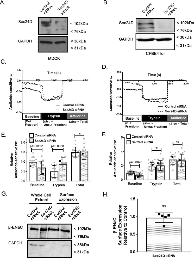
Sec24D regulates ENaC cleavage in MDCK αβγ ENaC and CFBE41o− epithelial cells. MDCK αβγ-ENaC cells (A, C, E, G, and H) or CFBE41o− epithelial cells (B, D, and F) were transiently transfected with nontargeted (control) or Sec24D siRNA and grown as polarized monolayers. Cells were mounted in Ussing chambers for Isc measurements (A–F) or underwent surface biotinylation (G and H) as described under “Experimental procedures.” A and B, representative immunoblot analysis performed after completion of the Isc measurements confirming siRNA-mediated depletion of Sec24D in MDCK αβγ-ENaC cells (A) or CFBE41o− epithelial cells (B). Shown are representative Isc traces from experiments in MDCK αβγ-ENaC (C) or CFBE41o− (D) cells, with annotations depicting experimental protocol. E and F, summary of amiloride-sensitive Isc measurements (relative to control baseline) are presented as mean ± S.D. (error bars); individual data points are depicted in gray, whereas representative traces from C and D are shown in black in E and F, respectively. Baseline Isc represents ENaC that is at the membrane in a cleaved/active form. E, application of trypsin to the apical surface in MDCK αβγ-ENaC cells acutely activates uncleaved/nearly silent ENaC (n = 13, p = 0.0122 for baseline, p = 0.0088 for trypsin, p = ns for total). Boldface points in E denote the representative Isc traces from C. F, Isc measurements of Sec24D-depleted CFBE41o− epithelial cells demonstrating decreased baseline ENaC-mediated Isc (n = 19, p = 0.0026). Boldface points in F denote the representative Isc traces from D. G, representative experiment demonstrating unchanged apical surface expression of β-ENaC by surface biotinylation. F, densitometric quantification of β-ENaC surface expression relative to control (n = 5 independent experiments, p = ns by Wilcoxon signed-rank test).

