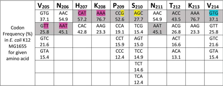
Figure 8.
Summary of codon usage in the internal initiation region of the eNISTmAb. The amino acid residues (205–214) from the eNISTmAb internal initiation region are shown. The possible codons for each amino acid are provided in descending order according to their relative occurrence in E. coli K12 MG1655 RefSeq coding DNA sequences (NCBI:txid511145 analyzed using HIVE-CUTs (30)). The codons used for eNISTmAb expression are shaded; the translation initiation motifs resulting from these codons are highlighted in pink and yellow for the putative S1-binding site and weak SD sequence, respectively. The internal initiation codon (GTG) is shown in cyan.

