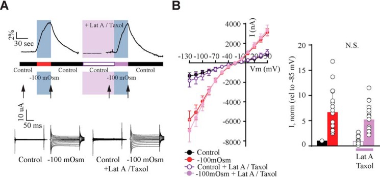
Figure 3.
Cytoskeletal rearrangements are not required for activation of TRPV4. A, experimental paradigm and representative volume traces (top). Application of a hyposmotic gradient is indicated by a blue bar, and drug application is shown by a pink bar. Representative current traces (bottom) were recorded as indicated by arrows (in control and hyposmotic solutions before drug application, after recovery and after latrunculin A and taxol application). B, summarized I/V curves with control solution (black), hyposmotic solution (red), control solution (white), and hyposmotic solution (light purple) after latrunculin A/taxol application. Insets, TRPV4-mediated current activity at −85 mV obtained after exposure to −100 mosm (red), in control solution with latrunculin A/taxol (white) or in hyposmotic solution with latrunculin A/taxol (light purple) was normalized to that obtained in control condition. N.S., not significant (p > 0.05); one-way ANOVA, n = 12 oocytes. Error bars, S.D.

