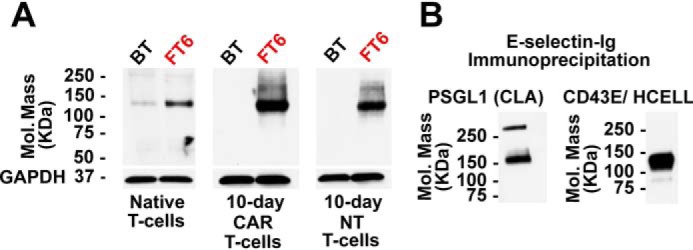
Figure 3.

Identification of the proteins carrying sLeX in exofucosylated CAR T-cells. A, Western blot using E-selectin–Ig chimera as a probe of BT- or FT6-treated native T-cells, 10-day CAR T-cells, and 10-day NT T-cells. GAPDH staining serves as a loading control. B, Western blot analysis of E-selectin–reactive glycoproteins of exofucosylated CAR T-cells. E-selectin–Ig immunoprecipitated glycoproteins from FT6-exofucosylated CAR T-cells were stained with antibodies against PSGL1 (left), and CD43 and CD44 (right). The numbers on the left indicate molecular mass in kilodaltons.
