Figure 1.
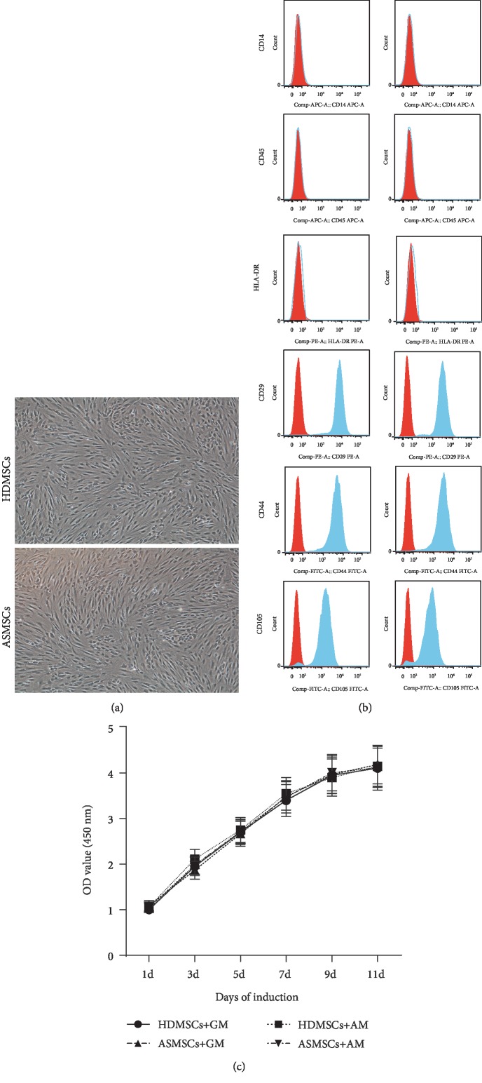
HDMSCs and ASMSCs exhibited similar morphologies, phenotypes, and proliferation rates. (a) HDMSCs and ASMSCs were both spindle-shaped, plastic-adherent cells. (b) HDMSCs (n = 30) and ASMSCs (n = 25) were negative for CD14, CD45, and HLA-DR and positive for CD29, CD44, and CD105, indicating a typical MSC phenotype. (c) HDMSCs (n = 30) and ASMSCs (n = 25) displayed equal proliferation capacities when cultured in either GM or AM from 1 to 11 days. The optical density (OD) values shown in (c) are presented as the means ± SDs.

