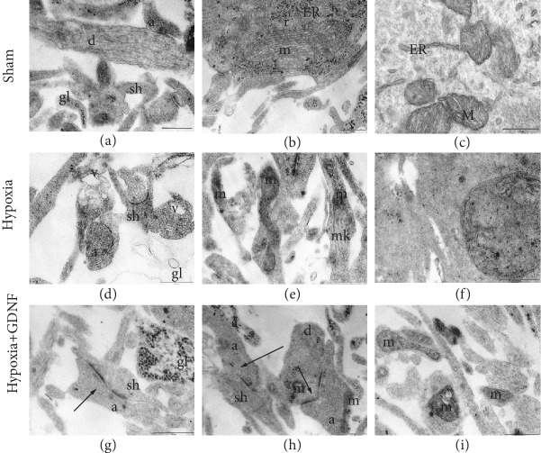
Figure 1.
Representative electron microscopy images of dissociated hippocampal cells on the day after acute normobaric hypoxia modelling. (а–c) Sham, (d–f) Hypoxia, and (g–i) Hypoxia+GDNF. (а) Axo-spiny and axo-dendritic asymmetric synapses. Mitochondria in the postsynaptic terminal of an intact structure and vesicles in an axonal bud have equal size and osmiophility, and glial outgrowths are filled with osmiophilic granules. (b) Axo-dendritic synapse. Mitochondria have moderate osmiophility, many cristae, and ribosomes, including in the endoplasmic reticulum. (c) Mitochondria in a cell body and ribosomes, including in the endoplasmic reticulum. (d) Axo-spiny asymmetric contacts with a concave surface, vacuoles from destroyed mitochondria in the outgrowth, and a shell from the empty glial outgrowth. (e) Mitochondria in a neuronal outgrowth with an irregular form and osmiophilic vesicles with additional membranes among synaptic vesicles are visible in a single axon. (f) Impaired mitochondrion in a cell body; the internal structure is completely disrupted. (g) Axo-spiny asymmetric perforated contact and glial outgrowth with osmiophilic granules. (h) Axo-spiny and axo-dendritic perforated contacts and mitochondria in the axon have an irregular form. (i) Mitochondria with modified structures in different outgrowths. а: axon; v: vacuoles; gl: glial outgrowth; ER: granular endoplasmic reticulum; d: dendrite; m: mitochondria; r: ribosomes; sh: spine; black arrow: mature chemical synapse. Scale bar: 0.5 μm.

