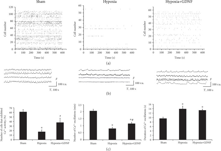
Figure 4.
Features of spontaneous calcium activity in primary hippocampal cultures on day 7 of the posthypoxic period. (a) Raster diagrams of spontaneous Ca2+ activity. The moments of oscillations occurrence are presented as strokes. (b) Representative recordings of spontaneous Ca2+ oscillations,. F: fluorescence intensity (relative units (r.u.)); T: time (seconds). (c) Main parameters of spontaneous calcium activity in primary hippocampal cultures: (left) proportion of cells exhibiting calcium activity; (middle) number of Ca2+ oscillations per min; (right) duration of Ca2+ oscillations. The data represent the mean values ± SEMs from three independent experiments. Statistical significance was calculated by one-way ANOVA; p < 0.05; ∗versus “Sham”; #versus “Hypoxia”.

