Figure 2.
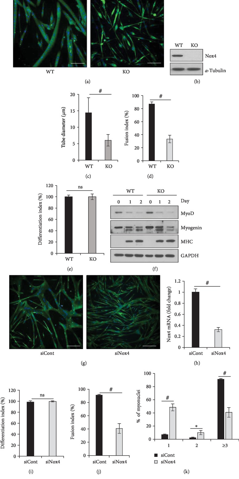
Nox4 is required for myoblast fusion of skeletal muscle. (a) Immunofluorescence images after primary myoblasts derived from WT and Nox4-KO mice were cultured in DM for 48 h and stained against MHC (green) and with DAPI for the nucleus (blue). Scale bar, 50 μm. (b) Nox4 expression was analyzed by immunoblotting. (c) Diameters of myotubes were measured using Nikon software. #p < 0.001 (n = 3). (d) Fusion index values for the experiments done in a were calculated as the percentage of nuclei in MHC-positive myotubes containing ≥3 nuclei among total nuclei in MHC-positive myotubes. #p < 0.001 (n = 3). (e) Differentiation index values for the experiments in a were quantified as the percentage of nuclei in MHC-positive cells among total nuclei. (f) The expression of MyoD, myogenin, and MHC proteins during differentiation was analyzed by immunoblotting in WT and Nox4-KO primary myoblasts. (g) Immunofluorescence images of primary myotubes cultured in DM for 48 h after cells were transfected with siCont or siNox4 and strained for MHC (green) and with DAPI (blue). Scale bar, 50 μm. (h) Nox4 mRNA levels as evaluated by RT-qPCR, using 36B4 for normalization. #p < 0.001 (n = 3). (i) Fusion index values for the experiments done in g were calculated as the percentage of nuclei in MHC-positive myotubes containing ≥3 nuclei among total nuclei within MHC-positive myotubes. #p < 0.001 (n = 3). (j) Differentiation index values for the experiments in g were quantified as the percentage of nuclei in MHC-positive cells among total nuclei. #p < 0.001 (n = 3). (k) Percentages of nuclei present in MHC-positive myotubes with the indicated number of nuclei were calculated in the experiment done in g. ∗p < 0.05, #p < 0.001 (n = 3). ns, no significant difference.

