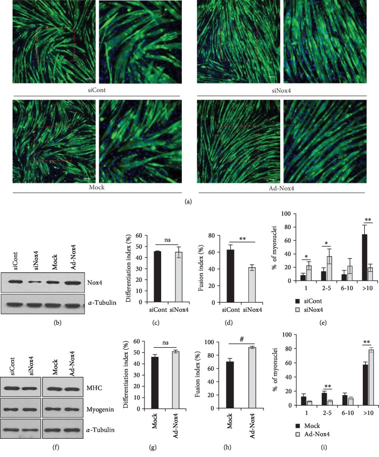
Figure 3.
Nox4 enhances myoblast fusion in C2C12 cells. (a) Immunofluorescence images of Nox4-KD or -overexpressing C2C12 cells cultured in DM for 3 days after staining for MHC (green) and with DAPI (blue). Scale bar, 50 μm. (b) Protein levels of Nox4 as analyzed by immunoblotting in Nox4-KD or -overexpressing C2C12 cells. (c) Differentiation index values were calculated as the percentage of nuclei in MHC-positive cells to total nuclei in Nox4-KD cells (siCont and siNox4) (n = 3). (d) Fusion index values were calculated as the percentage of nuclei present in MHC-positive myotubes (≥10 nuclei) to total nuclei in MHC-positive myotubes among Nox4-KD cells. ∗∗p < 0.01 (n = 3). (e) Percentages of nuclei in MHC-positive myotubes with the indicated number of nuclei were calculated in Nox4-KD cells. ∗p < 0.05, ∗∗p < 0.01 (n = 3). (f) Protein levels of MHC and myogenin as analyzed by immunoblotting in Nox4-KD or -overexpressing C2C12 cells. (g) Differentiation index values were calculated as the percentage of nuclei in MHC-positive cells to total nuclei in Nox4-overexpressing cells (mock and Ad-Nox4) (n = 3). (h) Fusion index values were calculated as the percentage of nuclei present in MHC-positive myotubes (≥10 nuclei) to total nuclei in MHC-positive myotubes among Nox4-overexpressing cells. #p < 0.001 (n = 3). (i) Percentages of nuclei in MHC-positive myotubes with the indicated number of nuclei were calculated in Nox4-overexpressing cells. ∗∗p < 0.01 (n = 3). ns, no significant difference.

