
Figure 2.
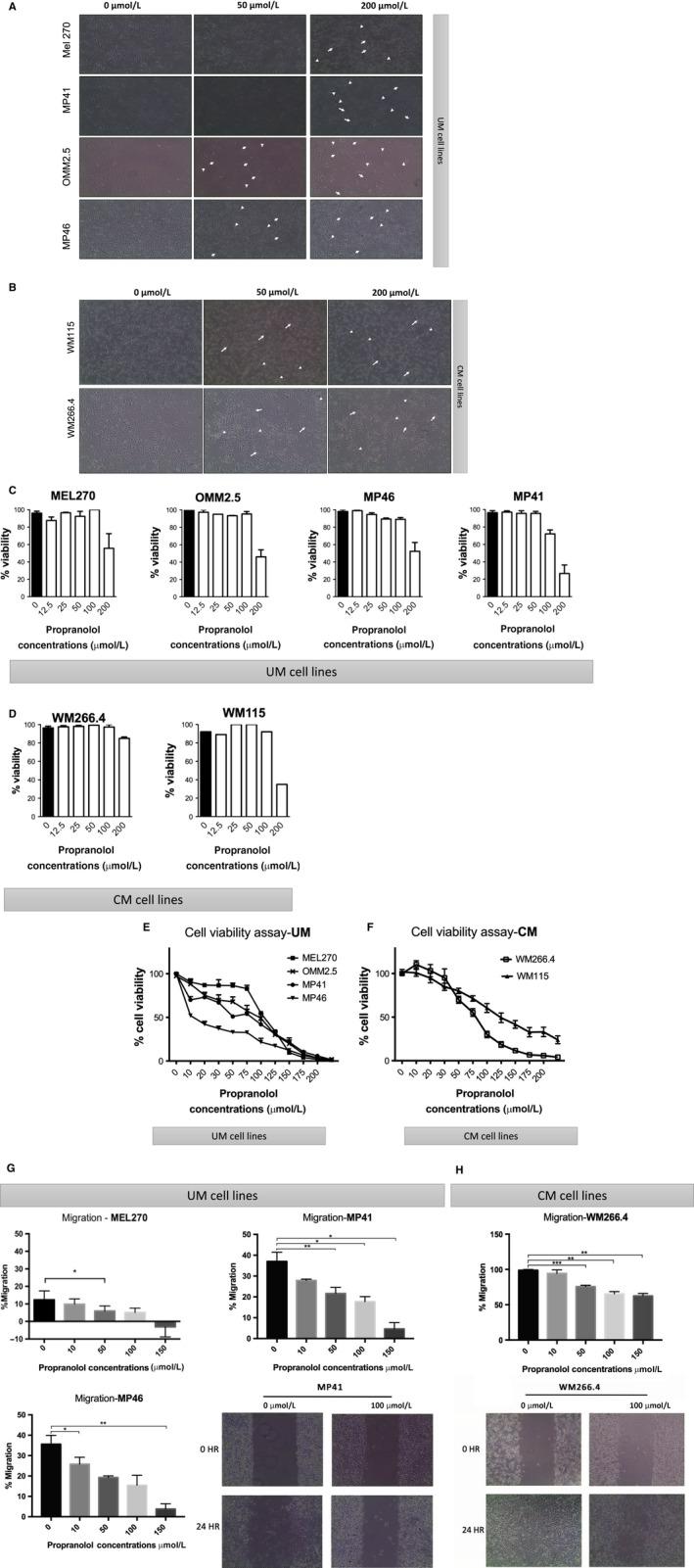
Effect of propranolol in uveal melanoma (UM) and cutaneous melanoma (CM) primary and metastatic cell lines. Representative images of cellular shape changes under 50 and 200 μmol/L propranolol treatment in (A) UM cell lines and (B) CM lines. 20× objective. Viability is shown by tryptan blue exclusion assay in triplicates in UM (C) and CM (D) cells. Cytotoxicity of propranolol at different concentrations in (E) UM and (F) CM cell lines is shown using CCK8 viability assay. Relative cell viability % was calculated as compared with the 0 μmol/L (no drug) control (100% viability). Error bar shows ± SD. Dose‐dependent inhibitory effects of propranolol on the percentage of migration of (G) UM (MP41, MP46, MEL270) and (H) CM (WM266.4 and WM115) cell lines were detected by wound healing assay after 24 h of drug exposure. *P < .05 vs control (0 μmol/L). Representative images of UM (MP41) and CM (WM266.4) migration assays (under phase contrast, 4× objective) are shown