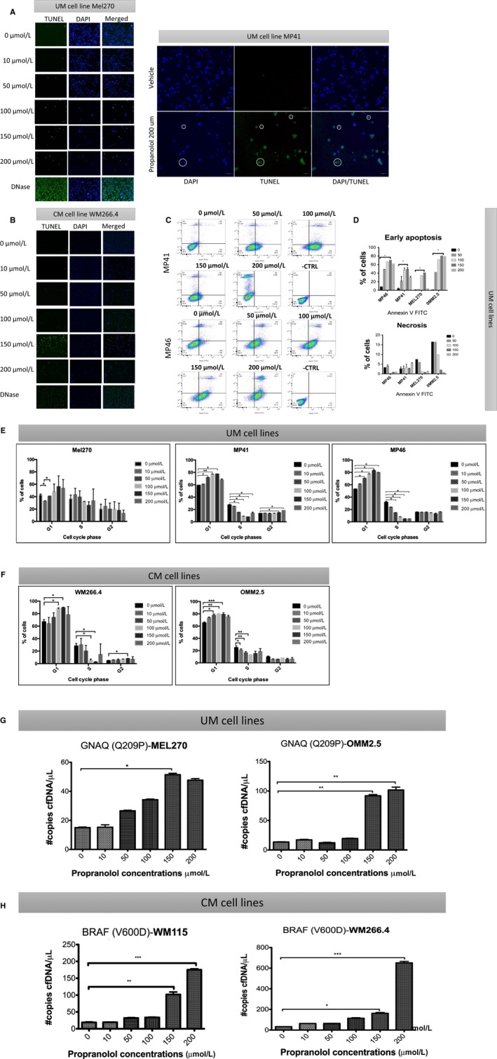
Figure 3.

The effect of propranolol on apoptotic cell death and cell cycle arrest in melanoma cells. TUNEL staining showed the presence of apoptotic cells under 50, 100, 150, and 200 μmol/L propranolol exposure during 24 h in (A) uveal melanoma (UM) (Mel 270 and MP41) and (B) cutaneous melanoma (CM) (WM266.4) cells. Cells under DNase were used as a positive control. Green color indicates TUNEL‐positivity, blue color marks nucleus. 20× objective. Representative flow cytometry analysis of UM using Annexin V FITC (C). Bar graph of early apoptosis, necrosis, and cell death on cultured UM cells (D). Cell cycle analysis was done by flow cytometry following propranolol exposure at 6 concentrations in UM (E) and CM (F) cell lines. Error bars represent ± 1 SD. *P < .05 vs control (0 μmol/L). cell‐free DNA (cfDNA) in the supernatant was assessed following 24 h treatment with propranolol in (G) UM (Mel 270 and OMM2.5) and (H) CM (WM266.4 and WM115) cell lines. The specific mutation for each cell line is indicated. *P < .05 vs control (0 μmol/L)
