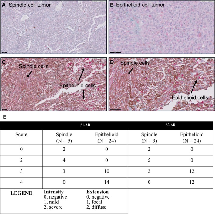
Figure 5.

Expression of β‐adrenoceptors in clinical UM samples. A, Spindle cell tumor showing focal areas of immunostaining (intensity 1, extension 1 = grade 2). B, Epithelioid cell tumor showing diffuse and intense staining (intensity 2, extension 2 = grade 4). (C, D) Same tumor with areas of spindle cell staining and epithelioid cell staining. Immunostaining was done using a red chromophore to differentiate from melanin pigment (brown). Scale 50 μm
