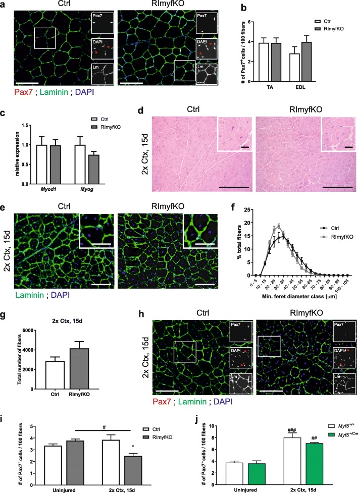
Fig. 3.
MuSC number after repeated injury is reduced in RImyfKO muscle. a Merged and single (insets) staining against Pax7 (red), laminin (green), and for DAPI (blue) of muscle cross-sections from 5-month-old Ctrl and RImyfKO mice. Red arrowheads in insets indicate DAPI-positive nuclei that are also Pax7-positive. Scale bar, 100 μm. b The number of Pax7-positive cells per 100 myofibers was counted in TA and EDL muscles from 5-month-old Ctrl and RImyfKO mice (n = 4–5). c Relative expression of transcripts encoding MyoD and MyoG in TA muscle from 5-month-old Ctrl and RImyfKO mice, normalized to Gapdh (n = 4). d–j Muscle regeneration was studied by two rounds of injections of cardiotoxin (Ctx) into TA muscle, 15 days post-injury (2x Ctx, 15 days). d H&E staining of TA muscle. Muscle fibers are regenerating irrespective of the genotype as indicated by the centralized nuclei. Scale bar, 100 μm and 10 μm (inset). e Immunostaining against laminin (green) and DAPI staining of TA cross-sections. Scale bar, 100 μm and 50 μm (inset). f Fiber size distribution and g total number of myofibers (n = 3). h Merged and single (insets) staining against Pax7 (red), laminin (green), and for DAPI (blue) of muscle cross-sections. Red arrowheads in insets indicate DAPI-positive nuclei that are also Pax7-positive. Scale bar, 100 μm. i Quantification of the number of Pax7-positive cells/100 myofibers of uninjured and injured muscle (n = 4). j Quantification of the number of Pax7-positive cells/100 myofibers in uninjured and injured muscle from Myf5+/+ and Myf5+/Cre mice (n = 4–5). The difference in the absolute number of Pax7-positive cells might be due to the different genetic background of the mice. Data represent mean ± SEM. *p < 0.05, Student’s t test (difference between genotypes). #p < 0.05, ##p < 0.01, ###p < 0.001, Sidak’s multiple comparisons test (difference between uninjured and injured muscles)

