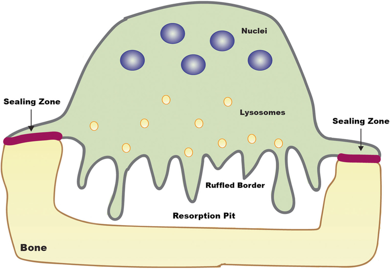
Fig. 1.
Cartoon illustrating acid-mediated extracellular proteolysis of bone by osteoclasts. A resorption pit comparable to an extracellular lysosome is created between the ruffled border of a multi-nucleated osteoclast and underlying bone. Sealing zones formed by podosome belts isolate the resorption pit. Lysosomes move toward the ruffled border and fuse with the membrane resulting in release of the cysteine protease cathepsin K into the resorption pit and incorporation of the vacuolar H+ ATPase in the lysosomal membrane into the ruffled border membrane. Acidification of the resorptive pit occurs as a result of generation of H+ ions by carbonic anhydrase II, also present in the ruffled border, and secretion of H+ ions into the pit by the vacuolar H+-ATPase. The organic matrix of the bone is degraded by the secreted cathepsin K

