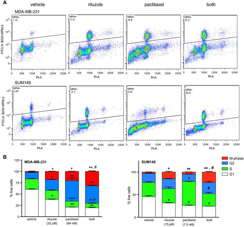
Fig. 6.
Riluzole and paclitaxel together enhance M-phase arrest in TNBC cells. MDA-MB-231 and SUM149 cells were treated overnight with riluzole and/or paclitaxel and the number of cells in M-phase, G2, S, or G1 were determined by FACS analysis. a Representative scatter plots of live cells staining positive for MPM-2 proteins (PI and FITC positive), proteins that are phosphorylated by M-phase promoting factor during mitosis. Riluzole increases and number of cells in M-phase for both MDA-MB-231 and SUM149 cells and riluzole and paclitaxel together (both) induce a higher percentage of cells expressing MPM-2 proteins compared to their respective vehicle control or paclitaxel-treated cells. b The data generated from the scatterplots above and from standard propidium iodide DNA staining were analyzed and graphed using prism software. G2 is calculated by subtracting percent of cells in M-phase from the G2/M values. The results are expressed as the percent of total live cells and are the average mean ± SEM of two experiments, performed in duplicate where *p < 0.05 and **p < 0.01 compared to vehicle control cells and #p < 0.05 compared to paclitaxel treatment alone. Riluzole significantly increases the percentage of cells in M-phase compared to vehicle-treated alone in both cell types and the percent of cells in M-phase is significantly greater in the combined treatment groups compared to paclitaxel treatment alone

