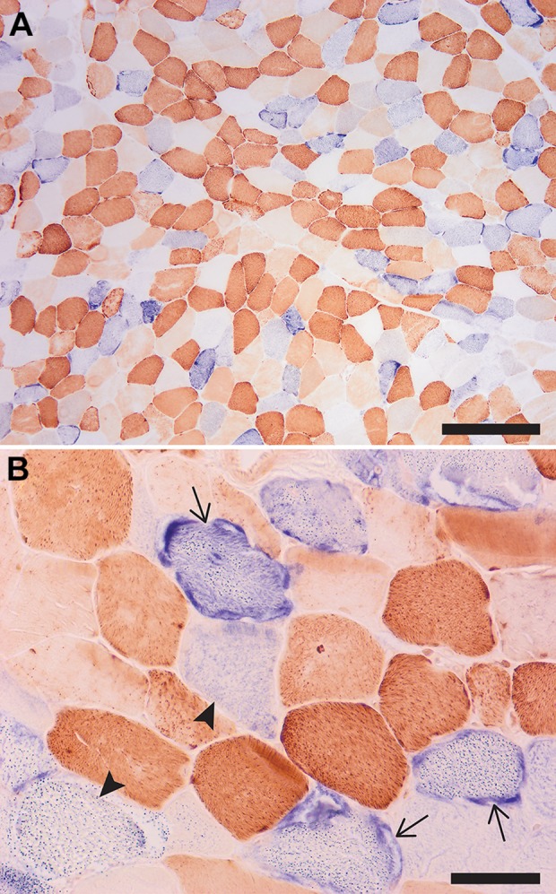
Figure 2.

Low (A) and high (B) magnifications of a cryosection sequentially stained with COX (brown) and SDH (blue) enzyme histochemistry. The fibers with abnormal mitochondria are staining either dark blue (COX-negative fibers with mitochondrial hyperplasia, which correspond to ragged red fibers; arrows) or light blue (COX-negative fibers without mitochondrial hyperplasia; arrowheads), while fibers depleted of mitochondria lack both COX and SDH activity and appear white/optically clear. Note the mosaic pattern of affected and unaffected fibers; in a normal muscle, only dark brown (type 1) and light brown (type 2) fibers would be seen. Scale bars: (A) 500 µm and (B) 100 µm. COX indicates cytochrome C oxidase; SDH, succinate dehydrogenase.
