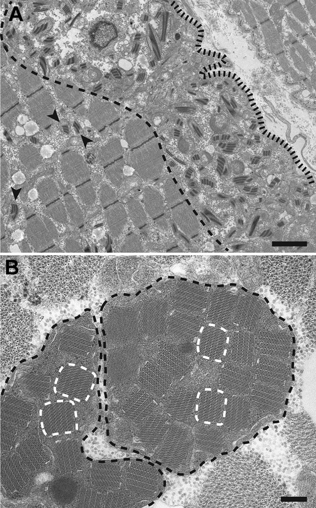
Figure 3.

A, Low magnification electron micrograph from a longitudinal muscle section showing a subsarcolemmal collection of abnormal mitochondria, which demonstrate a significant variation in the size and shape and contain “railway track”-like crystalline inclusions. (The subsarcolemmal zone is found between the sarcolemma [delineated by a black fence-like line] and the edge of the myofibrillar apparatus [delineated by a black dashed line]). The abnormal mitochondria are mostly found within this subsarcolemmal area, but can also be seen between the myofibrils (arrowheads). B, High-magnification electron micrograph from a muscle cross section shows 2 giant mitochondria (outlined by the black dashed lines) that are distended by crystalline inclusions with a “parking lot” appearance, a few of which are delineated by the white dashed lines. Scale bars: (A) 2 µm and (B) 0.2 µm.
